- Home
- Common Foot Problems
- Accessory Navicular Pain
Accessory Navicular
Written By: Chloe Wilson BSc(Hons) Physiotherapy
Reviewed By: FPE Medical Review Board
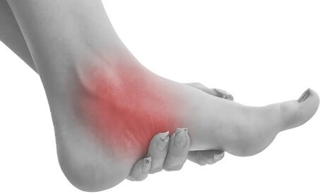
An accessory navicular is a small, pea-sized extra piece of bone found in the inner foot.
Only around 10% of the population have this extra bone and in most cases, it doesn’t cause any problems.
But if the bone or the surrounding tendon get irritated and inflamed it can lead to accessory navicular syndrome which causes pain on the inner side of the foot.
In most cases, navicular bone pain will settle down within a few weeks with correct treatment but more persistent cases may require surgery.
Here we will look at the common causes and symptoms of accessory navicular bone problems, how they are diagnosed and the best treatment options.
What Is An Accessory Navicular?
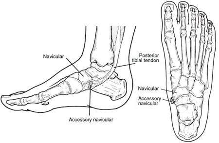
An accessory navicular is an extra piece of cartilage or bone found in the medial foot arch on the inner side of the foot, next to the navicular.
It usually sits within the posterior tibial tendon, one of the main stabilising muscles of the foot that supports the inner foot arch.
Only around 10% of the population have an accessory navicular, aka os tibiale externum or os navicularum. The cause of the extra bones is unknown but there is thought to be a genetic link.
Accessory navicular bones are congenital, meaning they are present from birth. They start off as a small piece of cartilage and, during adolescence, calcify into a hard lump of bone. People with accessory navicular bones typically have them in both feet, but they can be unilateral.
Accessory Navicular Types
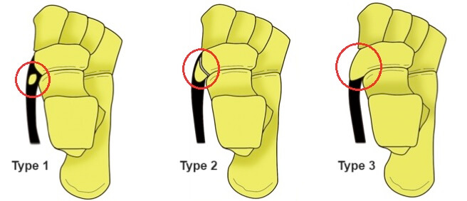
There are three different types of accessory navicular:
- Type 1 Accessory Navicular Bone: a small round or oval sesamoid bone that sits within the posterior tibial tendon. It is not connected to the navicular bone. Around 30% of accessory navicular bones are type 1. AKA os tibiale externum or os naviculare secundarium
- Type 2 Accessory Navicular: a heart-shaped or triangular bone measuring around 12mm in diameter. It is connected to the navicular bone by a thick layer of cartilage. Around 55% of accessory navicular bones are type 2. AKA prehallux or bifurcate hallux
- Type 3 Accessory Navicular: the largest type where the accessory navicular fuses (joins) to the navicular bone via a bony bridge, forming a cornuate (horn-shaped) bone. Around 15% of accessory navicular are type 3. AKA cornuate navicular
Causes Of Accessory Navicular Syndrome
In many cases, accessory navicular bones go completely unnoticed. But sometimes the accessory navicular and surrounding soft tissues can become irritated and inflamed, resulting in accessory navicular syndrome. This is most common with type 2 and type 3 accessory naviculars.
Common causes of accessory navicular syndrome include:
- Trauma: sudden trauma or injury to the foot, such as a forceful impact or a sprained ankle, can lead to irritation of the accessory navicular
- Footwear: wearing shoes that do not provide proper arch support can exacerbate foot mechanics issues, putting additional stress on the accessory navicular. Tight footwear may rub against the extra bone fragment causing chronic irritation and inflammation resulting in accessory navicular syndrome
- Overuse: activities that place repetitive stress on the foot, e.g. running, jumping or prolonged standing can irritate the accessory navicular and surrounding structures resulting in irritation and inflammation
- Biomechanics: individuals with altered foot biomechanics e.g. flat feet may be more prone to developing accessory navicular syndrome. These biomechanical factors can alter the distribution of forces in the foot, placing more strain on the posterior tibial tendon, impacting the accessory navicular
- Muscle Tightness: tightness in the posterior tibial muscle can result in increased tension and inflammation in the posterior tibial tendon which may irritate the accessory navicular
- Adolescence: accessory navicular pain often develops during adolescence due to a combination of ossification (where the accessory navicular changes from cartilage to bone) and increased activity levels
- Race: The prevalence of an accessory navicular has been found to be substantially higher in the Asian population, affecting around 50%
- Gender: Accessory navicular syndrome is approximately twice as common in females as males
Symptoms Of Os Navicularum
In most cases, accessory navicular bones are asymptomatic i.e. don’t cause any symptoms. But if the bone or surrounding tendon gets irritated, then accessory navicular syndrome can develop causing pain.
Common symptoms of accessory navicular syndrome include:
- Lump: there may a bony prominence i.e. small lump on the inner side of the foot, towards the top of the medial foot arch. You may be able to see or feel the lump, or both!
- Redness & Inflammation: there may be some swelling and redness around the accessory navicular lump on the inner side of the foot
- Pain: accessory navicular pain tends to be focused around the medial foot arch causing pain on the inside of the foot. People usually describe it as a dull or throbbing pain that is usually worse during or following activity. Occasionally, there may be sharp medial arch pain if there is direct pressure through the accessory navicular, or sudden twisting of the foot
- Weakness: you may find it difficult to push up onto your tiptoes if there is irritation of the posterior tibial from the accessory navicular
- Discomfort With Footwear: if the bony prominence rubs against your footwear, it can become quite uncomfortable
Diagnosing Accessory Navicular
In order to accurately diagnose accessory navicular bone problems, you should see your healthcare provider.
They will start by taking a history, asking you about your symptoms, how long you’ve had them, how they started and so on.
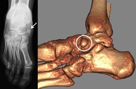
They will then examine your foot looking for any areas of swelling or tenderness, muscle strength and length, joint motion and foot function.
They may then send you for imaging studies, usually an x-ray, CT or MRI, to confirm the presence of an accessory navicular as well as rule out other possible causes.
Differential Diagnosis
There are a few other conditions that can cause inner foot pain that is similar to accessory navicular syndrome:
- Metatarsal Stress Fractures: a small break in one or more of the metatarsal bones
- Posterior Tibial Tendonitis: inflammation of the posterior tibial tendon often goes hand in hand with accessory navicular syndrome
- Tarsal Tunnel Syndrome: compression of the posterior tibial nerve at the inner ankle
- Tarsal Coalition: where a bony bridge forms between two of the foot bones, most typically the navicular and calcaneus
Accessory Navicular Bone Treatment
Accessory navicular syndrome treatment aims to reduce the symptoms of pain and inflammation and correct any underlying biomechanical issues. Accessory navicular treatment may involve:
- Rest: avoid any activities that exacerbate your symptoms such as high-impact exercises or activities that place excessive stress on the foot
- Immobilization: in more severe cases, your foot may be put in a cast or removable walking boot for a period of time, usually 2-3 weeks, to allow the affected area to rest and heal
- Ice: regularly applying an ice pack can help to reduce accessory navicular pain and inflammation. Find out how to safely and effectively use ice treatment
- Medication: your doctor may recommend non-steroidal anti-inflammatories (NSAIDs) such as ibuprofen to reduce accessory navicular bone pain and inflammation
- Physical Therapy: a rehab program is an important part of accessory navicular treatment and will usually involve a combination of strengthening, stretching and stability exercises for the foot and ankle. Accessory navicular syndrome exercises help to improve the strength and flexibility of the foot to reduce the strain on the extra bone which reduces the risk of recurrence of accessory navicular syndrome. Your physio can also provide advice on modifying activities and gait re-education
- Orthotics: special shoe insert orthotics for accessory navicular syndrome can help support the foot arches, improve foot mechanics and provide cushioning to reduce the pressure through the bone and tension on the tibialis posterior tendon
- Footwear: wear wide, cushioned, supportive shoes that do not rub on the accessory navicular to maintain proper alignment and reduce the risk of irritation of the bone
- Steroid Injection: your doctor may recommend a corticosteroid injection to reduce accessory navicular bone pain and inflammation, however this is not done routinely as it can cause temporary weakening of the tendon
In most cases, these non-surgical treatments are effective for treating accessory navicular syndrome and symptoms will usually settle down within a few weeks. It is important to continue with your exercise program and using any recommended orthotics to prevent recurrence.
Accessory Navicular Bone Surgery
If conservative treatment has been ineffective, or if symptoms keep recurring, then accessory navicular bone surgery may be recommended. There are a few different options depending on the type of accessory navicular and any associated foot issues:
- Accessory Navicular Excision: The most common type of surgery performed is an accessory navicular bone removal, aka excision. An incision is made on the inside of the foot and the accessory navicular bone is removed.
- Kinder Procedure: An incision is made just above the instep of the foot. The posterior tibial tendon is detached from the accessory navicular. The accessory navicular is separated from the navicular bone and the extra bone is removed. The surgeon then reattaches the posterior tibial tendon to the navicular bone.
- Realignment or Reconstruction Surgery: If there are biomechanical issues, realignment surgery may be done at the same time. This may involve releasing tight structures e.g. gastrocnemius calf muscle, tightening the tibialis posterior tendon or flat foot reconstruction where some of the foot bones are cut and repositioned with metal plates or screws.
Following accessory navicular bone surgery your ankle will be put in a cast or boot and you will be given crutches as you may be limited in how much weight you can put through the ankle for a few weeks. Accessory navicular bone surgery is successful in approximately 95% of cases.
Accessory Navicular Summary
An accessory navicular bone is a small extra sesamoid bone in the inner foot that sits within the posterior tibial tendon.
Also known as os navicularum or os tibiale externum, it is present from birth as cartilage and hardens (ossifies) during adolescence.
In most cases an accessory navicular does not cause any problems but if it gets irritated through overuse, muscle imbalance, altered foot biomechanics or friction it can become inflamed, causing accessory navicular syndrome and medial arch pain.
Accessory navicular bone treatment involves a combination of rest, ice, medication, physical therapy, exercises, orthotics and steroid injections.
If symptoms fail to settle or keep recurring, then accessory navicular bone surgery may be advised.
You may also be interested in the following articles:
- Inner Foot Pain
- Foot Arch Pain
- Ball Of Foot Pain
- Pain On Top Of The Foot
- Foot Lumps & Bumps
- Sharp Toe Pain
Page Last Updated: 11/16/23
Next Review Due: 11/16/25
Related Articles
Foot & Ankle Exercises
September 29, 2022
Diagnosis Chart
November 2, 2023
Burning Foot
March 8th, 2023
References
- Radiopaedia: Accessory Navicular. H. Knipe, May 2023
- ASM Journal: Prevalence And Classification Of Accessory Navicular Bone: A Medical Record Review. 2022
