- Home
- Anatomy Guide
Foot and Ankle Anatomy
Written By: Chloe Wilson BSc(Hons) Physiotherapy
Reviewed By: FPE Medical Review Board
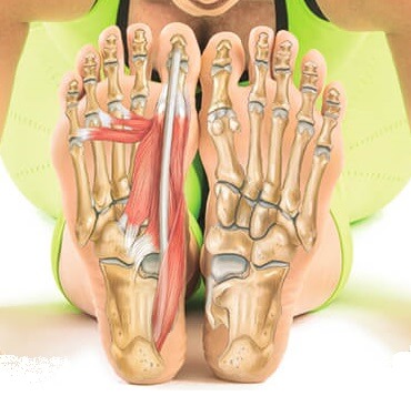
Foot and ankle anatomy consists of 33 bones, 26 joints and over a hundred muscles, ligaments and tendons.
This complex network of structures fit and work together to bear weight, allow movement and provide a stable base for us to stand and move on.
The foot needs to be strong and stable to support us, yet flexible to allow all sorts of complex movements with activities such as walking, running, jumping and kicking.
There are lots of things that can go wrong with the foot causing problems such as pain, weakness and instability.
Structures Of The Foot
When thinking about ankle anatomy, it helps to think about each of the different structures, how they fit together and what can go wrong. The main structures around the foot and ankle are:
- Bones: Provide the structure for the foot, comprising of lots of small joints to allow for small, subtle adjustments to help with balance
- Muscles: Soft tissues that contract and relax in groups so the foot can move properly with everyday activities such as walking, running and standing
- Ligaments: Strong connective tissues that hold the bones together and provide stability
- Tendons: Thick, cord-like structures that connect muscles to bone
- Joints: There are number of different joints in the foot which allow smooth, fluid movements in a range of directions
Let's have a look at each of these different elements of foot and ankle anatomy, how they fit together and what can go wrong.
1. Foot Bones
When thinking about foot and ankle anatomy, we usually divide the foot bones into three categories: the hindfoot, midfoot and forefoot.
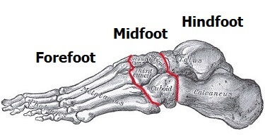
- The Hindfoot: the hindfoot comprises of the ankle joint, found at the bottom of the leg. This is where the ends of the shin bones, the tibia and fibula, meet the talus. Underneath this is the heel bone, aka the calcaneus.
- The Midfoot: The five bones of the midfoot are what make up our foot arches. They are arranged in a pyramid shape to be the shock absorbers of the feet. There is the navicular, cuboid and three cuneiform bones in the midfoot.
- The Forefoot: There are nineteen bones in the forefoot. The five metatarsals connect the midfoot to the toes and fourteen phalanges make up the toes themselves.
Common problems that arise in the foot bones include:
- Stress Fractures: small breaks in the bone usually from repetitive overloading in sports
- Hammer, Mallet & Claw Toe: Deformities at the toe joints that put the toes out of alignment. Affects 10-15% of the population
- Turf Toe: Hyperextension injury of the big toe - common in athletes. Also known as a toe joint sprain
- Bone Spurs: Excess bone growth in response to injuries, soft tissue tension or repetitive friction causing a hard lump
- Bunions: Gradually increasing deviation of the big or little toe, usually from tight-footwear that results in a large, painful lump on the side of the toe
Any damage that occurs to the foot and ankle bones is likely to affect any activity when you are on your feet and can cause issues further up the leg.
For more information about the different bones and joints and how they work together, visit the foot bones section.
2. Foot Muscles
Another important part of foot anatomy is the muscles. There are more than twenty muscles in the foot and they are commonly divided into two groups:
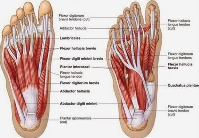
- The Intrinsics: The muscles that originate in the foot itself, on either the top (dorsum) or base (plantar) aspect of the foot
- The Extrinsics: The muscles that originate from the front and back of the lower leg and attach into the foot such as the calf muscles, gastrocnemius and soleus.
Muscles work in pairs. One muscle relaxes and lengthens while the other contracts and shortens,. This allows smooth, controlled movement. The muscles are arranged in layers and are responsible for maintaining the correct shape of the foot for example the foot arches. They attach to the foot bones via tendons.
Weakness and tightness in the calf and foot muscles not only cause foot pain but are also a common cause of knee, hip and back pain. Stretches and strengthening exercises can make a big difference.
The most common problem affecting the foot muscles is tendonitis. This is where there is inflammation and degeneration of the tendons, the cord part of the muscle where it attaches to the bone. Find out more in the foot tendonitis section.
3. Foot & Ankle Ligaments
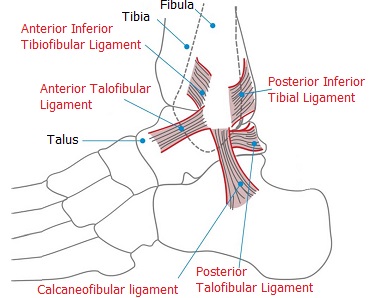
Another important part of foot and ankle anatomy are the ligaments.
Ligaments are strong, thick fibrous bands that connect bone to bone and hold them together. They are extremely important as they are the primary stabilizers of the ankle.
There are eleven ligaments around the ankle, connecting the various different bones of the hindfoot and midfoot. They work together to control all the different movements in the foot and ankle.
The most common ankle ligament injury is a ligament sprain, most commonly of the lateral ligament, aka anterior talofibular ligament. If an ankle sprain is not treated properly, it can cause long-term pain and instability in the ankle and foot as well as secondary problems such as cuboid syndrome which often goes undiagnosed.
4. Foot & Ankle Tendons
Another important part of foot and ankle anatomy are the tendons. Tendons are the thick cord-like structures that attach muscles to bone.
They transmit the force from the muscle to the bone causing the joint to move. So as the muscle contracts, it pulls on the tendon, which in turn pulls on the bone to move the joint. Tendons also help provide some stability to the foot.
If the ankle tendons are overloaded or overstretched they may become inflamed or even tear leading to tendonitis, tendonosis or rupture. The location of the pain will depend on which tendon is damaged.
The main tendons that are affected by foot tendonitis are:
- Peroneal Tendonitis: Causes pain down the back and outer side of the ankle/foot
- Posterior Tibial Tendonitis: Causes pain on the inner side of the foot
- Anterior Tibial Tendonitis: Causes pain across the front of the ankle
- Extensor Tendonitis: Causes pain along the top of the foot
- Achilles Tendonitis: Causes pain in the lower calf near the heel
- Sesamoiditis: Causes pain under the big toe
Find out more about the common causes, symptoms, diagnosis and treatment of these different types of foot tendonitis or how the tendons fit in with foot and ankle anatomy.
5. Ankle Joints
There are two joints that make up ankle anatomy:
- Ankle Joint: A synovial hinge joint between the tibia, fibula and talus bones. Allows the up and down movement at the ankle
- Subtalar Joint: A synovial plane joint between the talus and the calcaneus bones. Allows the side to side movement at the ankle.
There are then numerous joints between the different foot bones, held together by various ligaments.
Movements At The Ankle
Ankle anatomy allows a great deal of movement of the foot in different directions. The main movements of the foot are:
- Dorsiflexion: Where the foot bends up towards the shin
- Plantarflexion: where the foot and toes point downwards
- Inversion: where the foot twists inwards
- Eversion: where the foot twists outwards
Each of these movements is controlled by a complex combination of activity in the muscles, bones, ligaments and tendons.
Common Ankle Anatomy Problems
There are lots of different things that can go wrong with ankle anatomy that can cause foot problems. There may be weakness or tightness in the muscles, inflammation or degeneration in the tendons, damage or wear and tear to the bones.
Common foot and ankle problems include:
- Ligament Injuries: e.g. ankle sprain
- Foot Tendonitis: can affect any of the tendons
- Foot Lumps & Bumps: e.g. pockets of swelling or growths
- Bone Damage: e.g. fractures or mal-alignment
If you want help working out what is causing your pain, visit the foot pain diagnosis section.
You may also be interested in the following articles:
- Pain On Top Of Foot
- Foot Arch Pain
- Nerve Pain In The Foot
- Foot & Ankle Stretches
- Swollen Feet & Ankles
- Foot Numbness
If you want to find out more information about foot and ankle anatomy, continue through the following articles:
Bones | Muscles | Tendons | Ligaments
Related Articles
Page Last Updated: 19th November, 2024
Next Review Due: 19th November, 2026
